 首页
首页 400-620-6333


计算溶液所需的质量、体积或浓度。
这是演示店铺,请务下单付款,避免造成你的财物损失。
为了获得访问"阿拉丁铁蛋"实时聊天框的流畅支持体验,建议您使用Chrome浏览器或选择360浏览器极速模式(如何切换极速模式?),感谢您选择我们!
| 货号 (SKU) | 包装规格 | 是否现货 | 价格 | 数量 |
|---|---|---|---|---|
| C414608-250μg |
250μg |
期货  |
|
| 产品名称 | C3b |
|---|---|
| 规格或纯度 | 1.0 mg/mL,0.22 µm filtered |
| 产品介绍 |
Protein Purity >90% by SDS-PAGE Extinction Coeff. A280 nm = 1.03 at 1.0 mg/mL Molecular Weight 176,000 Da (2 chains) General Description C3b is derived from native C3 upon cleavage and release of C3a. It is prepared by cleavage with the alternative pathway C3 convertase. This is important because only a single bond in C3 is cleaved by the native convertase, while cleavage by other proteases, such as trypsin, results in multiple cleavages at many other sites in the protein. Native human C3b is a glycosylated (~2.8%) polypeptide containing two disulfide-linked chains. C3b is central to the function of all three pathways of complement (Law, S.K.A. and Reid, K.B.M. (1995)). Initiation of each pathway generates proteolytic enzyme complexes (C3 convertases) which are bound to the target surface. These enzymes cleave a peptide bond in C3 releasing the anaphylatoxin C3a and activating C3b. For a brief time (~60 µs) this nascent C3b is capable of reacting with and covalently coupling to hydroxyl groups on the target surface. Carbohydrates are the favored target, but protein hydroxyls and amino groups also react. This process of tagging the target surface with C3b is called opsonization. The reactive site in nascent C3b is a thioester (Tack B.J., et al. (1980); Pangburn M.K. and Müller-Eberhard H.J. (1980)) and C3b is linked to the target through a covalent ester bond (an amide bond is formed if C3b is attached to amino groups). Most of the C3b generated during complement activation never attaches to the surface because its thioester reacts with water forming fluid phase C3b which is rapidly inactivated by factors H and I forming iC3b. Surface-bound C3b is necessary in all three pathways for efficient activation of C5 and formation of C5b-9 complexes that lyse the target cell membrane. Surface-bound C3b and its breakdown products iC3b and C3d are recognized by numerous receptors on lymphoid and phagocytic cells which use the C3b ligand to stimulate antigen presentation to cells of the adaptive immune system. The end result is an expansion of target-specific B-cell and T-cell populations. Physical Characteristics & Structure Molecular weight: 176,000 daltons composed of two disulfide linked chains. The alpha prime chain (α’-chain) is 101,000 daltons (it contains the C3d domain) and the beta chain is 75,000 daltons. Alpha prime and beta chains are linked through a single disulfide bond. The pI of C3b is approx. 5.7.Upon cleavage of C3 by C3 convertases, C3a (77 amino acid fragment, 9083 Da) is released from the N-terminal of the alpha chain and C3b (176,000 Da) becomes attached covalently to the surface of the activator. The crystal-derived structures of both C3 and C3b have been described (Gros, P. (2008)) and these show that large conformational changes occur in the C3b portion of C3 following cleavage of the C3aC3b peptide bond. Function C3b alone has no enzymatic activity. It is a structural component of the alternative pathway C3 convertase (C3b,Bb), a structural component of the C5 convertases of all three pathways of complement activation (C3b,C3b,Bb and C3b,C4b,C2a) and a ligand for complement receptors CR1 and CR2.C3b is essential for effective complement activation and subsequent presentation of antigens to the cells of the adaptive immune system (Lambris, J.D. (1988)). Following recognition of a target, complement is activated by one of the three complement activation pathways and enzymes (C3 convertases) are formed on the target’s surface. These enzymes (C4b,C2a or C3b,Bb) cleave C3 after Arg 77 of the alpha chain releasing the anaphylatoxin C3a and depositing C3b on the target surface. Although there is a very weak C3 bypass system that operates through the classical and lectin pathways (C4b,C2a can activate C5 without C3b at ~1/2000 the rate of C3b,C4b,C2a), C3b is generallynecessary for effective C5 activation. Assays Assays of function include measurement of binding to factor B, factor H, factor P (properdin), C5 and cleavage by the protease factor I in the presence of the cofactor factor H. The later is the most convenient assays since it only requires factor H and I and SDS gels to analyze the cleavage of the alpha chain of C3b in 67,000 and 43,000 Da fragments. In vivo C3b arises from the proteolytic cleavage of C3. The serum concentration of C3 is 1.0 to 1.5 mg/mL with the average of 1.2 mg/mL which makes C3 the most abundant complement protein in blood. It represents approx. 2.5% of the total protein in blood and excluding albumin and immunoglobulins it is ~8% of the protein present in plasma. The primary site of synthesis is the liver, but C3 is also made in macrophages, neutrophils, astrocytes, and in endothelial and epithelial cells in many tissues of the body. During aggressive complement activation (in sepsis and at sites of infection) high concentrations of C3b may be formed, much of it fluid phase C3b. Regulation C3b is regulated by both fluid phase and membrane-bound inactivators. Factor I is a serine protease that can cleave C3b at two closely spaced locations. A single cleavage causes a structural rearrangement in C3b forming iC3b (inactive C3b) and iC3b lacks most of the binding sites that C3b possessed (for factor B, factor H, factor P, andC5). Cleavage and inactivation of C3b by factor I requires that a cofactor be bound to C3b (Pangburn M, et al. (1977)). The primary fluid phase cofactor is factor H (500 µg/mL in plasma). Some cell membranes, such as human erythrocytes, possess CR1 which can act as a cofactor for factor I. Most human cells and tissues have MCP (membrane cofactor protein) which also acts as a cofactor for factor I. CR1 only acts on C3b on cells or immune complexes other than the cell bearing the CR1 while MCP only acts on C3b attached to the cell membrane bearing the MCP. In the absence of factor I the interactions of C3b with factor H and CR1 inhibit C3b complement functions through competition with binding of factor B and through decay acceleration of the C3 convertase C3b,Bb. DAF (decay accelerating factor) is another membrane-bound protein that is present on most human cells and it interacts with C3b. It is not a cofactor for factor I and only promotes the dissociation of C3/C5 convertases containing C3b (C3b,Bb and C3b,C3b,Bb). The interactions of CR1, MCP, factor H, and DAF with C3b do not inactive the C3b itself and it is capable of continuing all of its complement functions once dissociated from them so long as it has not been cleaved by factor I. Genetics Human chromosome location of C3 gene 19p13.3-p13.2. Mouse chromosome location chromosome 17 and rat chromosome 9. Accession numbers K02765 (human) and K02782 (mouse). Human C3 genomic structure: the gene spans 41 kb with 41 exons Deficiencies Complete human C3 deficiencies are rare but a number of cases have been found. Importantly, adults with this condition have been found so although there is a high risk due to impaired immunity, it is not necessarily fatal. A well characterized case of a deficient two year old child demonstrated that the deficiency is associated with recurrent pyogenic infections, impaired dendritic cell differentiation , impaired ability to acquire B cell memory and deficient regulatory T cell development. Vaccination produced only a small, short term antibody response (Ghannam A, et al. (2008)). Other human cases and numerous animal experimental models support these conclusions (Singer, L, et al., (1994)). The association of these immune system defects with C3 deficiency strongly supports a major role for C3 in innate and adaptive immune responses.The absence of C3 also results in failure to opsonize bacteria with C3b resulting in reduced phagocytosis, failure to release C3a and severely reduced ability to generate C5a and C5b which impairs generation of the terminal complement complex C5b-9. Diseases The deposition of C3, that is, the attachment of C3b to microorganisms or host tissues is the hallmark of complement activation at inflammatory sites. Many diseases exhibit histochemically identifiable C3b deposits as part of their pathology, or at least as markers of pathology (Law, S.K.A. and Reid, K.B.M. (1995); Ross, G.D. (1986)). These diseases include ischemia/reperfusion events such as heart attacks and strokes and bacterial, viral, parasitic and fungal infections. Antibody-mediated autoimmune diseases such as systemic lupus erythematosus, rheumatoid arthritis, and autoimmune hemolytic anemia are characterized by C3b deposition on tissues. Even when the antibody response is not directed at the host complement can be deposited. A major function of complement is to aid the macrophage phagocytic system in the removal of circulating immune complexes. High levels of complexes can overwhelm this system leading to the deposition of immune complexes and active complement. In tissues and in the kidney this leads to glomerulonephritis, dense deposit disease, and arthritis. C3b deposits also signal complement activation in diseases such as paroxysmal nocturnal hemoglobinuria, inherited hemolytic uremic syndrome, transplant rejection and inflammatory skin diseases such as angioedema. Precautions/Toxicity/Hazards The source of this protein is human serum, therefore precautions appropriate for handling any blood-derived product must be used even though the source was shown by certified tests to be negative for HBsAg, HTLV-I/II, STS, and for antibodies to HCV, HIV-1 and HIV-II. MSDS available upon request. |
| 来源 | Normal human serum (shown by certified tests to be negative for HBsAg and for antibodies to HCV, HIV-1 and HIV-II) |
| 物理外观 | Liquid |
|---|---|
| 储存缓冲液 | 10 mM sodium phosphate, 145 mM NaCl, pH 7.3 |
| 储存温度 | 避免反复冻融,-80℃储存 |
| 运输条件 | 超低温冰袋运输 |
输入批号以搜索COA:


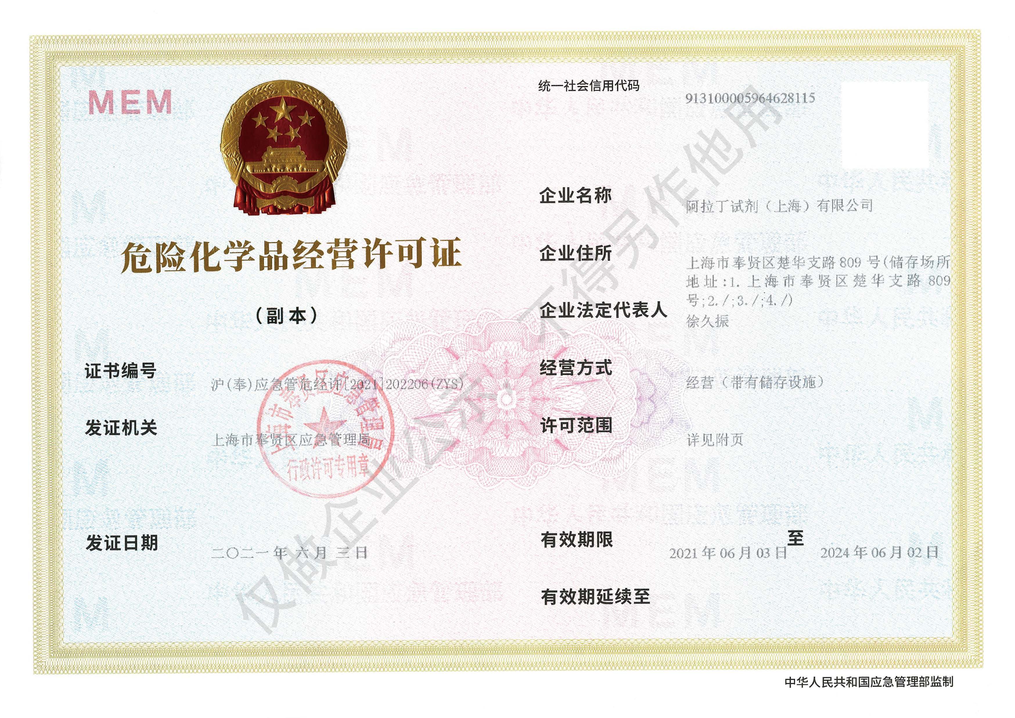 危险品化学品经营许可证(带存储)