这是演示店铺,请务下单付款,避免造成你的财物损失。
 首页
首页 400-620-6333
liposomes
PRODUCT DESCRIPTION
The phospholipid bilayers are composed of phosphatidylcholine and cholesterol. All liposomal formulations are suspended in sterile phosphate buffered saline (PBS):
– 10 mM Na2HPO4;
– 10 mM NaH2PO4;
– 140 mM NaCl.
All our liposomes are slightly negatively charged and will therefore not clog together. The liposomes in the suspension are not sized, meaning that it contains smaller and larger liposomes as well (up to 3 micron). On average, the size of the liposomes are 1.7 micron.
Clodronate liposomes: a suspension of artificially prepared lipid vesicles encapsulating clodronate. The concentration of clodronate in the suspension is ca. 5 mg / mL. Clodronate is encapsulated in the liposomal vesicles in the form of CH2Na2Cl2O6P2 · 4 H2O.
PBS liposomes: a suspension of artificially prepared lipid vesicles encapsulating an aqueous PBS solution. These do not contain clodronate and can be used for control experiments.
Fluorescent DiI Liposomes: a suspension of artificially prepared lipid vesicles encapsulating an aqueous PBS solution, labelled with the fluorochrome DiI. These do not contain clodronate and can be used to investigate whether liposomes injected via a particular administration route are able to reach the macrophages to be studied.
STORAGE AND DIRECTIONS OF USE
Upon arrival, liposomes should be stored between 4 – 8 ºC (or 39 – 47 ºF). The liposomal suspensions should never be frozen, nor be exposed to extreme high temperatures. This can cause disturbances to the phospholipid bilayers, possibly leading to leakage of clodronate out of the liposome.
Before administration, let the liposomes reach room temperature first and gently shake or stir the suspension. Liposomes tend to precipitate after some time, causing an inhomogeneous distribution in the vial. When injection takes too much time, the liposomes may even precipitate in the syringe. If multiple animals are injected using the same syringe, this can cause differential dosing.
Dilution of the suspension is discouraged, but if necessary use PBS or saline.
We advise our customers to use our liposomal formulation within 16 weeks after shipment. Use after the expiry date has occurred is strongly discouraged. After this period the risk of contamination increases, and a slight loss of function could occur.
Macrophages play an important role in immune and non-immune defence mechanisms. They form a first line of defence against bacterial, viral and other forms of microbiological contamination penetrating into the bodies of vertebrates. Macrophages are large cells, found in almost all bodily tissues where they can have varying forms and names (e.g. Kupffer cells, alveolar macrophages, microglia, osteoclasts, red pulp macrophages). Macrophages “scavenge”, they ingest and digest all foreign substances, microbes, cancer cells and cellular debris that might be potential pathogens. This process is called phagocytosis. Macrophages further regulate functions of many non-phagocytic cells, mainly through mediation of soluble molecules such as cytokines and chemokines. They are involved in innate immunity, adaptive immunity and can have (anti-) inflammatory effects.
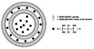
Liposomes are artificially prepared spheres and consist of concentric phospholipid bilayers. When phospholipids are dispersed in water, the hydrophilic heads will make up both outer parts of the liposome, whereas the hydrophobic fatty acid groups will make up the inner part (see figure 1).
Aqueous compartments separate the bilayers, and hydrophilic molecules can be dissolved in it, resulting in liposome-encapsulated molecules. Clodronate (dichloromethylene-bisphosphonate or Cl2MBP) is a hydrophilic molecule that can be encapsulated within phospholipid bilayers. Free clodronate does not easily cross cell membranes, and is rapidly cleared (i.e. within minutes) from circulation by the renal system. However, when entrapped in a liposome, the clodronate liposome is ingested by macrophages and cannot escape it (see figure 2).

The phospholipid bilayers are digested by lysosomal phospholipases, whereas clodronate is not digested and remains in the macrophage. The more phospholipid bilayers and liposomes are ingested by the macrophage, the more clodronate will accumulate within the macrophage. Exceeding a certain intracellular concentration, clodronate will eliminate the macrophage by initiating its programmed cell death, i.e. apoptosis.
Administration protocols
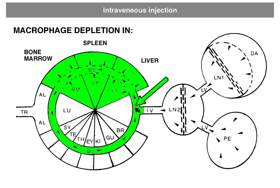
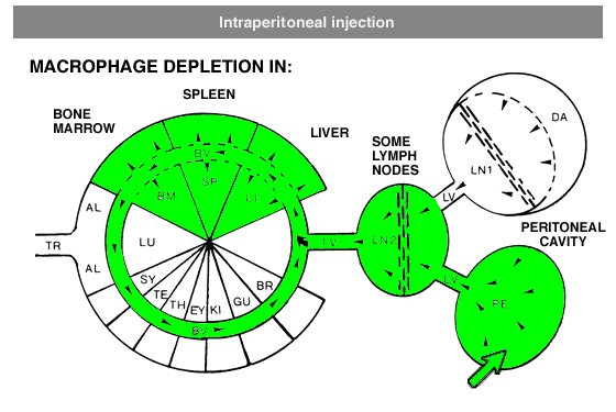
Administration protocols should be chosen carefully and depend on several factors, for instance: the type of macrophage you intend to deplete (e.g. Kupffer cells, alveolar macrophages, microglia, osteoclasts, red pulp macrophages), the time in which you intend to maintain depletion (e.g. short or long term), the (animal) model, and other experimental factors. In vitro application of clodronate liposomes is possible, albeit that they are specifically suitable to study macrophages in vivo. Below you can see schematic representations of the tissues and macrophages that can be reached through different administration routes. Please note that not all tissues and routes are represented here.
Abbreviations: AL = lung alveoli, BM = bone marrow, BR = brain, BV = blood vessels / circulation, DA = draining area of lymph node, EY = eye, GU = gut / intestines, IV = intravenous delivery, KI = kidney, LI = liver, LN = lymph node, LU = lung, LV = lymph vessels, PE = peritoneal cavity, SP = spleen, SY = synovial cavity in joint, TE = testis, TR = trachea.
In general, it is recommend to inject 100 µL of suspension / 10 grams of animal weight for intravenous injection. Raising the dosage considerably may lead to blockage of capillaries. Intravenous administration of clodronate liposomes will lead to maximum depletion of liver and spleen macrophages in ca. 24 hours. Dependent on the subset of macrophages they will remain depleted for ca. 5 days. After that time, new macrophages will replace the depleted ones: macrophage precursors, monocytes, that are formed in bone marrow and released in circulation will arrive at their destination and further differentiate into mature macrophages. Monocytes, macrophage precursors, can also be depleted. To prevent macrophage repopulation, multiple injections can be administered every 2-3 days to target monocytes. See for instance: Sunderkötter, C., Nikolic, T., Dillon, M. J., Van Rooijen, N., Stehling, M., Drevets, D. A., & Leenen, P. J. (2004). Subpopulations of mouse blood monocytes differ in maturation stage and inflammatory response. The Journal of Immunology, 172(7), 4410-4417.For intraperitoneal administration, the recommended injection dosis (i.e. 100 µL of suspension / 10 grams of animal weight) can be increased considerably. This route also depletes peritoneal macrophages, but will take longer to deplete macrophages in liver and spleen (ca. 3 days). Depletion is slower and more gradual, since the liposomes have to be carried from the peritoneal cavity to circulation by lymph flow via the thoracic duct, which is a passive form of transport.
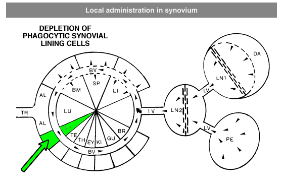
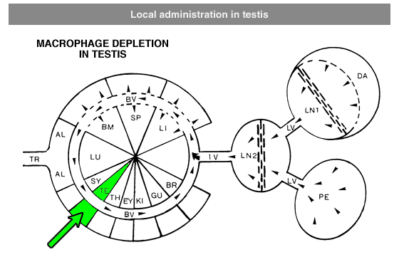

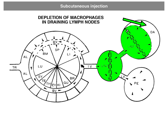
Local administration is often required to target macrophages that are difficult to reach, such as macrophages in testis or the phagocytic synovial lining cells.For depletion of alveolar macrophages, clodronate liposomes can be administered intranasally as well as intratracheally. The difference is that intranasal liposomes may be spoiled in the oesophagus if not administered properly, whereas intratracheal instillation will deliver all liposomes in the lung. Please note that these routes only target alveolar macrophages, and not interstitial macrophages. These can be targeted through depletion of blood monocytes in circulation. See for instance: Huang, L., Nazarova, E. V., Tan, S., Liu, Y., & Russell, D. G. (2018). Growth of Mycobacterium tuberculosis in vivo segregates with host macrophage metabolism and ontogeny. Journal of Experimental Medicine, 215(4), 1135-1152.For subcutaneous injection the maximum volume to be injected depends on the storage capacity of the injection site.
RESULTS
Sections of mouse liver, stained with monoclonal antibody F4/80 Normal liver with positive Kupffer cells
Liver of a mouse, 2 days after injection with clodronate-liposomes. Kupffer cells have been depleted

Sections of mouse spleen stained for acid phosphatase Normal spleen with positive splenic macrophages

Spleen of a mouse 2 days after injection with clodronate liposomes. Only few macrophages are left

REFERENCES
- First publication of the approach
Van Rooijen, N., & Van Nieuwmegen, R. (1984). Elimination of phagocytic cells in the spleen after intravenous injection of liposome-encapsulated dichloromethylene diphosphonate. Cell and tissue research, 238(2), 355-358.
- Application in research and therapeutics
van Rooijen, N., & van Kesteren-Hendrikx, E. (2002). Clodronate liposomes: perspectives in research and therapeutics. Journal of liposome research, 12(1-2), 81-94.
- Elimination and blocking of macrophages in general
Rooijen and Annemarie Sanders, N. (1997). Elimination, blocking, and activation of macrophages: three of a kind?. Journal of leukocyte biology, 62(6), 702-709.
- Mechanism macrophage apoptosis mediated by clodronate liposome
Van Rooijen, N., Sanders, A., & van den Berg, T. K. (1996). Apoptosis of macrophages induced by liposome-mediated intracellular delivery of clodronate and propamidine. Journal of immunological methods, 193(1), 93-99.
- Depletion of Kupffer cells in liver
Van Rooijen, N. I. C. O., & Sanders, A. (1996). Kupffer cell depletion by liposome‐delivered drugs: comparative activity of intracellular clodronate, propamidine, and ethylenediaminetetraacetic acid. Hepatology, 23(5), 1239-1243.
- Depletion of macrophages in spleen
Van Rooijen, N., Van Nieuwmegen, R., & Kamperdijk, E. W. A. (1985). Elimination of phagocytic cells in the spleen after intravenous injection of liposomeencapsulated dichloromethylene diphosphonate. Virchows Archiv B, 49(1), 375.
Van Rooijen, N., Kors, N., & Kraal, G. (1989). Macrophage subset repopulation in the spleen: differential kinetics after liposome‐mediated elimination. Journal of leukocyte biology, 45(2), 97-104.
Van Rooijen, N., Kors, N., Vd Ende, M., & Dijkstra, C. D. (1990). Depletion and repopulation of macrophages in spleen and liver of rat after intravenous treatment with liposome-encapsulated dichloromethylene diphosphonate. Cell and tissue research, 260(2), 215-222.
- Depletion of macrophages in lymph nodes
Delemarre, F. G. A., Kors, N., Kraal, G., & Van Rooijen, N. (1990). Repopulation of macrophages in popliteal lymph nodes of mice after liposome‐mediated depletion. Journal of leukocyte biology, 47(3), 251-257.
- Depletion of alveolar macrophages in lung
Thepen, T., Van Rooijen, N., & Kraal, G. (1989). Alveolar macrophage elimination in vivo is associated with an increase in pulmonary immune response in mice. Journal of Experimental Medicine, 170(2), 499-509.
- Depletion of peritoneal macrophages
Biewenga, J., van der Ende, M. B., Krist, L. F., Borst, A., Ghufron, M., & van Rooijen, N. (1995). Macrophage depletion in the rat after intraperitoneal administration of liposome-encapsulated clodronate: depletion kinetics and accelerated repopulation of peritoneal and omental macrophages by administration of Freund’s adjuvant. Cell and tissue research, 280(1), 189-196.
- Depletion of testis macrophages
Bergh, A., Damber, J. E., & Van Rooijen, N. (1993). Liposome-mediated macrophage depletion: an experimental approach to study the role of testicular macrophages in the rat. Journal of endocrinology, 136(3), 407-NP.
- Depletion of phagocytic synovial lining cells in joints
Blom, A. B., van Lent, P. L., Holthuysen, A. E., van der Kraan, P. M., Roth, J., van Rooijen, N., & van den Berg, W. B. (2004). Synovial lining macrophages mediate osteophyte formation during experimental osteoarthritis. Osteoarthritis and cartilage, 12(8), 627-635.
- Depletion of perivascular and meningeal macrophages in the central nervous system
Polfliet, M. M., Goede, P. H., van Kesteren-Hendrikx, E. M., van Rooijen, N., Dijkstra, C. D., & van den Berg, T. K. (2001). A method for the selective depletion of perivascular and meningeal macrophages in the central nervous system. Journal of neuroimmunology, 116(2), 188-195.
标签:
标签
订购
订单
货物签收
退换货
退货
售后
发票
易制毒
运输
储存
产品运输
产品储存
有效期
复测
复检
失效日期
复检日期
批次
日期信息
易制爆
易制爆品
易制爆化学品
纯度
纯度等级
试剂纯度
规格
纯度级别
grade
含量
纯度规格
等级
试剂等级
蛋白储存
抗体保存
抗体储存
蛋白保存
单位帐户
个人帐户
用户
注册
登录
付款
银行帐户
葡聚糖
Dextran
帐户
修改密码
重设密码
B2B对接
订单对接
产品对接
数据对接
电子采购集成
e-Commerce Solutions
eProcurement
发货
签收
配送方式
第三方快递
配送
运费
退款
脂质体
脂质粒
氯膦酸盐脂质体
CAS注册表号
纯度和质量
体外实验
细胞渗透性
看不到产品
产品外观
分子量和分子式
溶解
储存产品
溶解度
运输条件和产品标签
包装尺寸和称量准确度
巨噬细胞清除
氯膦酸盐(脂质体)的半衰期
物美价廉
产品可进行的实验类型
产品的SDS和分析证书
危化品
GHS
GHS 分类
积分
普鲁卡因胺标记
试剂盒
氰基硼氢化钠
还原剂
检测量
甲基硼烷
2-AB标记试剂盒
缓冲液
RNA
疾病研究
冻干肽的储存和使用
脂质体悬浮液储存
氯磷酸盐脂质体浓度
溶解和储存肽溶液
脂质体的大小和电荷
脂质体悬浮介质
制备氨基酸溶液
检测Dil标记的脂质体
培养基
Medium
唾液酸
糖蛋白
唾液酸定量分析
还原胺化反应
2-甲基吡啶-甲硼烷复合物
石墨化碳
介孔
介孔二氧化硅
官能化二氧化硅
介孔材料性能和应用
Graphitized Carbon
Mesoporous
Mesoporous Silica
Functionalized Silica
Properties and applications of mesoporous materials
透明质酸
liposomes
chlorophosphonate liposomes
CAS号
体内实验使用剂量
In vitro
cell-permeable
dissolve
store product
solubility
Conditions of carriage; Product labels
half-life of clodronate (liposomes)
Pack sizes and weighing accuracy
macrophage depletion
氯磷酸盐脂质体
(clodronate) liposomes
肽
氯磷酸盐脂质体注射
Cheap-and-fine
Types of experiments that can be performed on the product
SDS and analysis certificate of the product
聚合物
聚酰胺6
聚丙烯
Polymer
Polyamide 6
Polypropylene
发光聚合物
聚苯乙烯
聚(1,4-亚苯基)
聚芴
聚(噻吩)
聚喹啉
水溶性LEP
Light-Emitting Polymers
Polystyrene
poly(1,4-phenylene)
Polyfluorene
Poly(thiophene)
Polyquinoline
Water-soluble LEP
导电聚合物
聚苯胺
聚噻吩
Conductive polymer
Polyaniline
Polythiophene
Product appearance
Molecular weight and molecular formula
银纳米材料
用于生物学应用的银纳米材料
PCR
MIQE指南
技术支持
Technical Support
危险化学品
光催化
石墨烯
石墨烯在光催化中的应用
导电油墨
石墨烯油墨
石墨烯薄膜
纳米银颗粒分散液
印刷电子
全甲基化试剂盒
Buffer
N-异丙基丙烯酰胺
PNIPAM
细胞膜片组织工程
N-Isopropylacrylamide
Cell membrane tissue engineering
rewards points
膜剥离
免疫印迹
Western blotting
purity and quality
peptide
Chlorophosphate liposome injection
Storage and use of lyophilized peptides
store the liposomal suspension
concentration-of-the-clodronate-liposome-suspension
Dissolve and store peptide solution
size-and-charge-of-the-liposomes
Dissolved amino acids
detect-the-Dil-labeled-liposomes
MIQE Guidelines
Conductive ink
Graphene ink
Graphene film
Silver nanoparticle dispersion
Printed electronics
can't see the product
凝胶电泳
核酸
琼脂糖
聚丙烯酰胺
聚丙烯酰胺凝胶电泳
琼脂糖-丙烯酰胺复合凝胶
Gel electrophoresis
Nucleic acid
Agarose
Polyacrylamide
Polyacrylamide gel electrophoresis
Agarose acrylamide composite gel
硅
砷化镓
锗
砷化铟
Indium arsenide
Gallium arsenide
Silicon
Germanium
核苷
核苷酸
Nucleoside
Nucleotide
核酸电泳
Nucleic acid electrophoresis
聚乙二醇(PEG)
Polyethylene Glycol (PEG)
碳化硅
氧化磷
Silicon carbide
Phosphorus oxide
生物缓冲液
Biological buffer
富勒烯
Fullerene
聚乙二醇修饰剂
聚乙二醇酸
聚乙二醇NHS活性酯
PEG-COOH
PEG-NHS Ester
聚乙二醇胺
PEG-NH2
聚乙二醇酸(PEG-COOH)
有机溶剂法
水溶液法
巯基聚乙二醇
金纳米粒子
Polyglycolic acid
Aqueous solution method
organic solvent method
磷酸盐缓冲盐水
可溶于水的有机溶剂
聚乙二醇马来酰亚胺
洗涤液
马来酰亚胺储备液
polyethylene glycol maleimide
Maleimide stock solution
detergent
mercapto polyethylene glycol
Gold Nanoparticles
Polyethylene Glycol NHS Active Ester
Phosphate Buffered Saline
water soluble organic solvent
聚乙二醇五氟苯酚酯
Polypentafluorophenol
聚乙二醇醛(PEG-CHO)
烷氧胺
PEG-SPDP交联剂
Polyglycol aldehyde
alkoxyamine
聚乙二醇氨氧基
玻璃层析柱
Glass chromatographic column
透析袋
Dialysis bag
D101大孔树脂
联合碳化透析袋
层析柱
即用型透析袋
Ready to use dialysis bag
琼脂糖凝胶
即用型CE膜透析袋
单壁碳纳米管
Single walled carbon nanotubes
糖酵解
5-磷酸核糖
TCA循环
表皮生长因子
Epidermal growth factor
nucleotide
Glycolysis
Ribose 5-phosphate
TCA cycle
生物正交化学
叠氮炔烃点击化学
Bioorthogonal Chemistry
Azide-Alkyne Click Chemistry
新标签
材料科学
生物医学材料
Material science
Biomedical materials
抗体
Antibody
Antibodies
蛋白质
稳定性
protein
stability
内毒素
endotoxin
抗坏血酸
谷胱甘肽
硫辛酸
α-生育酚
BSA
HSA
蔗糖
ED50
细胞因子浓度
ascorbic acid
glutathione
Lipoic acid
α- tocopherol
核酸合成
cytokine concentration
Specific activity unit
International Unit
干扰素
GM-CSF
FGF
神经营养素
sucrose
interferon
neurotrophin
具体活动单位
国际活动单位
International Activity Unit
Nucleic acid synthesis
活性单位
电泳
剥离石墨
perylene
芘
卟啉
exfoliated graphite
Pyrene
Porphyrin
fullerenes
钙离子指示剂
离子载体
Calcium ion indicator
Ionic carrier
寡核苷酸
亚磷酰胺法
磷酸三酯法
Nucleotides
Oligonucleotides
Phosphoramidite method
Phosphate Triester Method
凝胶定量
聚丙烯酰胺凝胶
Agarose gel
Polyacrylamide gel
Gel quantification
糖基化
天冬氨酸
糖苷酶
蛋白酶
Glycosylation
aspartic acid
Glycosidase
protease
光电子
射频
发光二极管
光伏发电
GaAs
RF Devices
radio frequency
light emitting diode
PV
单糖分析
烯基含氟砌块
烯化反应
消除反应
交叉偶联反应
亲电氟化
二氧化钛
转换器
半导体材料
Alkenyl fluorinated building blocks
Olefination reactions
Elimination reactions
Cross-coupling reactions
Electrophilic fluorination
Titanium dioxide
converter
Semiconductor material
木瓜蛋白酶
治疗肿瘤
改善肉质嫩度
细胞分离操作
Reagent test kit
Detection amount
Monosaccharide Analysis
papain
treat tumors
Improve meat tenderness
Cell isolation procedure
直接带隙半导体
间接带隙半导体
多透明非晶氧化物半导体
如氧化铟镓锌
direct bandgap semiconductor
indirect bandgap semiconductor
indium gallium zinc oxide
Polytransparent amorphous oxide semiconductor
水凝胶的降解
酶促降解
光降解型水凝胶
Degradation of hydrogels
enzymatic degradation
photodegradable hydrogel
氟西汀
肟菌酯
索拉非尼
含氟砌块
Sorafenib tosylate
Fluoxetine
Trifloxystrobin
Fluorine block
2-甲基吡啶硼烷
V-Tag 糖肽标记
V-Tag Glycopeptide Labeling
外切糖苷酶清理板
外切糖苷酶净化板
Exoglycosidase Clean-up Plate
白血病
JAK家族激酶
leukemia
JAK family kinases
核酸酶
细胞和分子生物学研究
靶向效率
脱靶突变
Nuclease
Cell and Molecular Biology Research
Targeting efficiency
Off target mutation
木糖
单糖释放和标记试剂盒
乙醇
三氯乙烯
异丙醇
半导体制造
生物制药
酶催化
酶活性
光刻胶
光刻流程
Semiconductor manufacturing
Photoresist
Photolithography process
酶分析技术
Pharmaceuticals
Enzyme catalysis
enzymatic activity
Enzyme analysis technology
色谱柱
HPLC系统
标记聚糖
Chromatographic column
HPLC system
Labelled glycan
PEG-SPDP Crosslinker
polyethylene glycol aminooxy
D101 macroporous resin
Combined carbonization dialysis bag
Ready to use CE membrane dialysis bag
New Label
xylose
Monosaccharide Release and Labeling Kit
肿瘤细胞
抗体-药物偶联物
包封率
装载量
产率
胶束交联策略
胶束的稳定性
encapsulation efficiency
loading capacity
yield
Stability of micelles
Micellar crosslinking strategy
纳米粒子
多肽的释放
可注射水凝胶
release of polypeptides
Injectable hydrogel
HPLC
胃蛋白酶活性测定
血红蛋白
高效液相色谱法
交换柱
Exchange column
CEX纯化柱
CEX Cartridges for O-glycans
S滤芯
S Cartridges
T1滤芯
真空歧管
T1 Cartridges
Vacuum Manifold
净化板
Procainamide
Purification plate
tumor cell
Antibody drug conjugates
口服给药
nanocarrier
oral administration
纳米载体尺寸
氨基聚乙二醇化
质检证书
稳定性同位素
C13
N15
神经干细胞
NSCs
neural stem cell
胰蛋白酶
解离细胞
Trypsin
dissociated cell
本征半导体
非本征半导体
PD-1
PD-L1
癌症免疫抑制
Cancer immunosuppression
有机砌块
二氟甲基
Organic block
Difluoromethyl
预甲基化清理板
Pre-Permethylation Clean-up Plate
EB10 墨盒
EB10 Cartridges
EB10 滤芯
EC50 滤芯
EC50 Cartridges
砌块
Building Blocks
砌块的应用
砌块的常见类型
砌块的官能团种类
砌块的角色
常用的砌块
砌块产品的竞争壁垒
砌块的应用规模
砌块的合成技术
类器官
Organoids
DMSO-d6
D2O溶液
NMR核磁溶剂
NMR solvents
同位素溶剂
同位素
微量元素分析
6Li金属
稳定性同位素标记标准
氘代氯仿
NMR用溶剂
氘代试剂
锂离子电池
硅负极材料
Lithium-ion Batteries
Silicon Anode Materials
芳基氟化反应
Aryl fluorination
PAN凋亡
PAN apoptosis
氮杂吲哚
蛋白质组学
代谢研究
标签
阿拉丁产品标签
试剂标签
化学品标签
化学试剂标签
试剂标签格式
小分子
抑制剂
选择指南
Small molecule
inhibitor
Selection Guide
氟代烷基化
Togni试剂
隔膜
维生素D
维生素D3
维生素D2
酶
催化剂
生物催化技术
Enzyme
Catalyzer
Biocatalytic technology
Stable isotope
氢化硅
Silicon hydride
药物开发
定制化医学
生物标记
生化网络
Deuterium reagent
Solvent for NMR
Deuterated chloroform
细胞培养
芯硅谷
耗材
贴壁细胞
DNA
TC处理
细胞培养皿
悬浮细胞
细胞培养瓶
细胞冻存管炸裂
细胞冻存管
cell culture flask
cryogenic vials
离心管
Fries重排
路易斯酸
布朗斯特酸
碳水化合物代谢
脂质代谢
谷氨酰胺代谢
核苷酸代谢
Carbohydrate metabolism
Lipid metabolism
Glutamic acid metabolism
Nucleotide metabolism
Fries rearrangement
lewis acid
Brønsted acids
Proteomics
Metabolic research
傅-克酰基化反应
Friedel–Crafts Acylation
蛋白样品制备
无机催化剂
羟醛缩合反应
免疫沉淀
Immunoprecipitation
Catalytic agent
Inorganic catalyst
Aldol condensation reaction
重组蛋白
Recombinant proteins
重组蛋白复溶与保存
蛋白标签
Baeyer-Villiger氧化
生物缓冲液选择指南
label
Aladdin product label
Reagent label
Chemical label
Chemical reagent label
Reagent label format
卡尔费休试剂
过渡态理论
反应热
化学中间体
有机催化剂
过渡金属催化剂
Azaindoles
Transition-state theory
Heat of reaction
Chemical intermediate
Catalyst
Organic catalyst
Transition metal catalyst
酶探针
ELISA实验
Enzyme probes
ELISA formats
锂金属氟化电池
傅-克烷基化反应
Friedel-Crafts alkylation
Fluorinated block
Fluoroalkylation
Togni reagent
离子液体
苯并四咪唑
均苯并四咪唑
不对称催化
手性催化剂
丙二烯砌块
Allenes Building Blocks
有机化学
Knoevenagel缩合反应
Lithium ion battery
the diaphragm
NF-kB信号通路
NF-kB
RNA病毒
信号通路
NF-kB signaling pathway
Signaling pathway
RNA virus
Heck反应
Heck Reaction
卡尔费休法
卡尔费休样品
Karl Fischer Reagent
卡尔费休滴定法
库仑法
容量法
FAQ
常见问题解答
常见问题
激动剂
拮抗剂
使用说明
Antagonists
Frequently asked questions
Instructions
细胞因子
炎症
Cytokines
Inflammation
巨噬细胞
Biginelli反应
药物应用
Biginelli Reaction
Block
Drug Application
Karl Fischer titration
测试
ADC连接子
抗体偶联药物
ADC药物
连接子
可降解连接子
金催化剂
氢化用金催化剂
N-杂环卡宾配体
NHC
均相催化
卡宾
配体
MRI定位细菌
纳米颗粒
病原体检测和鉴定
Cannizzaro反应
Gold Catalysts
The Gold Catalyst for Hydrogenation
克莱门森还原反应
Clemmensen Reduction
cell culture
i-Quip
Consumables
Adherent cell
磁性颗粒
氧化铁磁性颗粒
金磁性颗粒
二氧化硅磁性颗粒
超顺磁性聚苯乙烯(SPP)颗粒
金纳米材料
碳纳米材料
硅纳米颗粒
磁性材料
磁分离
磁诊断
传感器
药物传递
靶向治疗
Wolff-Kishner还原反应
Wolff-Kishner Reduction
磺酰氯
磺酰胺
合成化学领域
药物化学领域
ADC linker
Antibody conjugated drugs
ADC drugs
Linker
Cleavable linkers
Sulfonyl Chlorides
Sulfonamides
Field of synthetic chemistry
Field of medicinal chemistry
MRI Localization of Bacteria
Applications of Nanoparticles
Pathogen Detection and Identification
Cannizzaro Reaction
靶点
Target
阿兹海默症
电解液
阻燃剂
正极材料
蛋白质分离
病原体检测
靶蛋白
异源二聚体
表面增强拉曼光谱(SERS)
纳米材料
二氧化硅
金纳米颗粒
光声成像
细胞跟踪
靶向给药
Surface-Enhanced Raman Spectroscopy (SERS)
SERS Nanomaterials
Silicon dioxide
Photoacoustic imaging
Cell tracking
Targeted drug delivery
SERS
白蛋白
Baeyer-Villiger Oxidation
叔丁基亚磺酰胺
Ellman's Sulfinamides
金鸡纳生物碱
Cinchona Alkaloids
奎宁
Asymmetric catalysis
Quinine
MNPs
四氧化三铁
细胞器分离
光催化剂
烯烃加氢
反马氏规则
加氢官能化
Ligand
Photocatalyst
Olefin hydrogenation
Anti-Markovnikov
Hydrofunctionalization
DNA分离
RNA分离
HBTM
Chiral catalyst
小分子药物
Nanoparticles
Magnetic material
Drug delivery
Small Molecule Drugs
光氧化还原
氧化还原
自由基
可见光
Transition metal catalysts
Photoredox
REDOX
Free radical
Visible light
NPs
疫苗递送
马氏规则
Markovnikov’s Rule
格氏试剂
Guidelines for the selection of biological buffers
神经元
神经传递
加氢催化剂
氢化
还原
Catalysts
Hydrogenation Catalysts
Hydrogenation
Reduce
原子转移自由基聚合
三氟甲基化
ATRP
Trifluoromethylation
Apply Store Credit
Purchas and Redeem Gift Card
IgG抗体
一抗
二抗
重组抗体
单克隆抗体
多克隆抗体
Order Status
Refund
Order
Organic chemistry
Knoevenagel Condensation Reaction
IgG
Primary antibody
Secondary antibody
Recombinant antibody
Monoclonal antibody
Polyclonal antibody
格氏反应
Grignard reaction
生长因子
转化生长因子β
骨形态发生蛋白-1
Order workflow
Growth factor
TGF-β
BMP-1
N-Heterocyclic Carbene Ligands
Homogeneous catalysis
Carbene
MPs
Iron Oxide Magnetic Particles
Gold Magnetic Particles
Silica Magnetic Particles
Superparamagnetic Polystyrene (SPP) Particles
Nanogold
Carbon Nanomaterials
Silicon Nanoparticles
Magnetic separation
Magnetic diagnosis
Sensor
Targeted therapy
Alzheimer's disease
electrolyte
Flame retardant
Cathode material
Protein Separation
Pathogen Detection
Target Protein
Heterodimer
Albumin
Ferroferric Oxide
Organelle separation
DNA separation
RNA isolation
Vaccine delivery
Grignard Reagent
Neurons
neurotransmission
磁性纳米颗粒
量子点
碳纳米管
nano carrier size
amino polyglycolation
Determination of pepsin activity
hemoglobin
COA
Intrinsic Semiconductor
Extrinsic Semiconductor
The applications of Building Blocks
The common types of drug Building Blocks
The substituted functional groups of the Building Blocks
The role of the block
Common Building Blocks
Barriers to competition of building block products
The application scale of molecular Building Blocks
The synthetic techniques of molecular Building Blocks
D2O solutions
Isotope solvent
Trace element analysis
Isotopes
6Li metal
Stable isotope-labeled standards
Vitamin D3
Vitamin D2
Vitamin D
Drug development
Customized medicine
Biomarker
Biochemical network
TC processing
cell culture dish
Suspension cells
细胞不贴壁
培养瓶/皿/板
Cell cryovial burst
centrifuge tube
电极材料
Protein sample preparation
Reconstitution and preservation of recombinant protein?
Protein tag
Lithium Metal Fluoride Batteries
Li-ion Batteries
Ionic liquids
固态可充电电池
Karl Fischer Method
coulometric method
volumetric method
狄尔斯–阿尔德反应
Macrophage
核酸递送
QDs
Quantum Dots
Nucleic Acid Delivery
TEMPO催化氧化
TEMPO Catalyzed Oxidations
Magnetic Nanoparticles
Carbon Nanotube
Gene Delivery
趋化因子
泛素
泛素受体
巨核细胞
白细胞介素
肿瘤坏死因子
Chemokines
Ubiquitin
Ubiquitin receptor
Megakaryocyte
Interleukin
TNF
Diels-Alder Reaction
胶体金
电子显微镜
单电子转移
交叉偶联
SWCNTs
咪唑
不对称
Organocatalyst
Asymmetric
咪唑烷酮
Imidazolidinone
脯氨酸
脯氨醇
杂环砌块
1,2,4-三氮唑衍
Heterocyclic Building Block
1,2,4-Triazole
Proline
Prolinol
溶胶凝胶法
气溶胶法
水热合成法
喷雾干燥法
Sol-Gel Method
Aerosol Method
Hydrothermal Synthesis Method
Spray Drying
SET
Cross-coupling
吖丁啶
杂环化合物
Heterocyclic block
Heterocyclic compound
醇氧化催化剂
TEMPO
AZADOL
手性磷酸
还原胺化
丙烯基化
烷基化
Alcohol oxidation catalyst
点击化学
BINOL
VAPOL
VANOL
不对称合成
Asymmetric synthesis
纳米晶体
荧光
半导体
OD
癌症检测
癌症治疗
癌细胞
体内成像
Cancer detection
Cancer treatment
Vivo imaging
石墨烯量子点
GQDs
可溶性并五苯前体
电子材料
OFET
Soluble Pentacene Precursors
Electronic Materials
神经肽
血管紧张素
阿尔兹海默症
高血压
Neuropeptide
Angiotensin
hypertensive
论文
文献提交
科研论文
期刊登记
论文登记
论文登记有奖
MSDS
SDS
TDS
PbS
硫化铅
钙钛矿
光伏
Lead sulfide
perovskite
photovoltaic
荧光探针
荧光染料
Fluorescence
Fuorescent Probe
Fluorescent dyes
Nanocrystalline
Semiconductor
Azetidine
PQD
钙钛矿量子点
半导体纳米晶体
空穴传输材料
Perovskite Quantum Dot
Semiconductor Nanocrystal
HTMs
1,3-噻唑砌块
1,3-Thiazole building blocks
染料聚集
Dye-Aggregation
免疫球蛋白
抗体亚型
重链和轻链
Immunoglobulin
Antibody isotype
Heavy chain and light chain
罗丹明
红移
蓝移
Rhodamine
Red shift
Blue shift
太阳能电池
Solar cell
有机发光
Organic light emitting
蛋白质印迹
内参抗体
Loading control
流式细胞术
Flow Cytometry
前向散射
侧向散射
二向色滤光片
多色流式细胞仪
Forward scatter
Side scatter
Dichroic filters/mirrors
The multicolor flow cytometer
基因敲除
免疫细胞化学
Knockout gene
ICC
有机发光二极管
有机光电电池
OLED
OPV
荧光团
直接/间接检测
酶标记物
Fluorophore
Direct/Indirect Detection
Enzyme markers
点击试剂
铜催化
叠氮化物
炔烃
CuAAC
Click Chemistry
Click Reagen
Copper catalyzed
azide
alkyne
参考物质
认证参考物质
分析标准品
分析证书
Reference material
Certified reference material
Analytical standard
Certificate of analysis
Graphene
graphene quantum dot
酶联免疫吸附
免疫组织化学
ELISA
WB
IHC
生物正交
叠氮化合物
四嗪
四唑
Click Reagent
Bioorthogonal
Azide Compound
Tetrazine
Tetrazole
Returns
Returns and Refunds
business accounts
personal accounts
account types
硝酮
Nitrone
SPANC
同种型对照
Isotype Control
查看更多


 危险品化学品经营许可证(带存储)